Live-cell imaging to overcome blind spots in long experiments
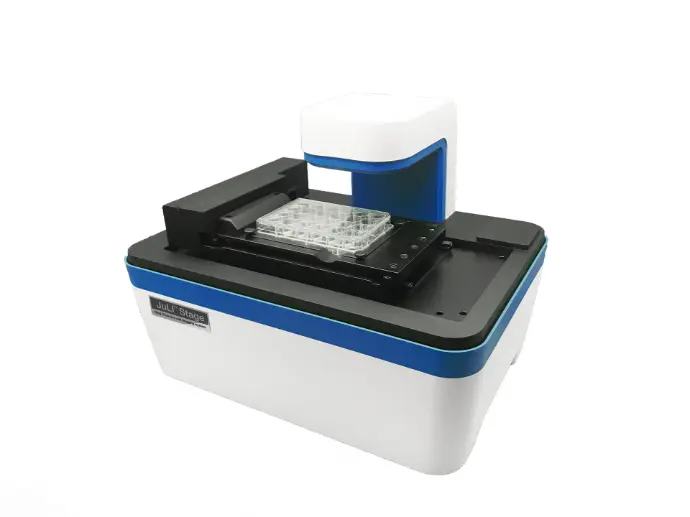
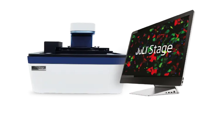
JuLI™ Stage is a real-time live cell imaging system designed to operate inside a standard CO₂ incubator enabling time-lapse monitoring without repeatedly removing cultures for microscopy
It combines automated multi-position imaging with
analysis options so you can record cell behavior over
time and generate visual evidence (images, time-lapse
sequences, growth trends) for better decisions during
culture
Advantages of the JuLI™ Stage live cell imaging system
Compact and incubatorcompatible.
Built to run in common incubator environments, enabling uninterrupted observation close to physiological conditions.
Automated scanning across
wells and positions.
A fully automated X-Y-Z stage supports multi-position imaging workflows for plates and other vessels - reducing manual microscope time.
Fluorescence + brightfield for
richer insight.
Supports multi-channel fluorescence imaging (e.g., GFP/RFP/DAPI) alongside brightfield, enabling monitoring of expression, morphology, and dynamics.
Key Features and Benefits
Time-lapse recording and “cell history”.
Captures time-lapse sequences to document what
happened during the full culture period - not just one
endpoint.
Broad vessel compatibility.
Designed to support common culture formats, including multi-well plates across a wide range of densities.
Software support and export.
Includes software workflows for acquisition and data handling, supporting practical documentation and sharing of results.